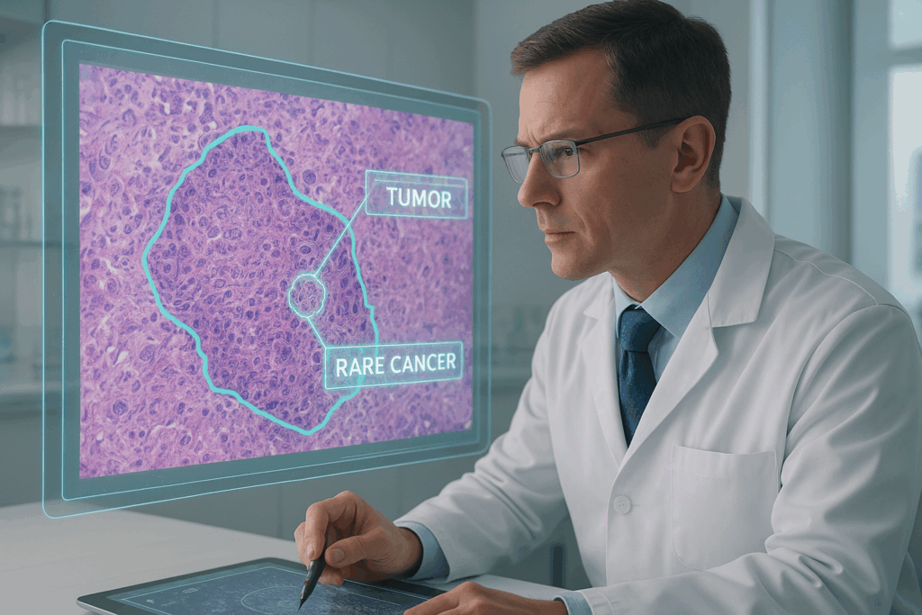
Rare cancers, such as sarcomas, pose a unique challenge for medical research and AI development. These underrepresented cancer types often lack sufficient high-quality annotations, making it difficult to train reliable AI models. When it comes to these complex cases, precision is not just important—it’s essential.
At the heart of building accurate AI for rare cancers lies a simple truth: quality annotation guided by pathology expertise makes all the difference.
Challenges in Rare Cancer AI Development
Developing AI solutions for rare cancers requires overcoming several hurdles:
- Limited exposure to atypical cases: Many rare cancers appear infrequently, meaning that every case must be carefully examined and annotated by specialists.
- Complex histological features: Rare cancers often display subtle variations in tissue structures, demanding consistent and precise labeling.
- Need for medical collaboration: To ensure clinical relevance, AI development must be closely aligned with expert pathologists’ insights.
How Marteck Solutions Supports Precision AI
At Marteck Solutions, we specialize in pathology-guided annotation and labeling services designed to meet the unique needs of rare cancer research. Our approach focuses on three core principles:
- Expert-led annotation of low-frequency cases: Our team works closely with experienced pathologists to ensure even the rarest cases are accurately represented.
- Consistent labeling standards: Every histological feature is labeled with precision, enabling reliable analysis and minimizing variability.
- Collaborative medical insights: We maintain close communication with clinical specialists, ensuring that every annotation aligns with real-world diagnostic standards.
By combining deep medical expertise with meticulous annotation practices, Marteck Solutions empowers researchers and developers to build AI solutions that are both accurate and clinically meaningful.
Looking ahead: With our pathology-driven annotation and labeling services, your next breakthrough in rare cancer AI is within reach.
Contact Marteck Solutions today to explore how we can support your AI initiatives with precision and medical rigor.