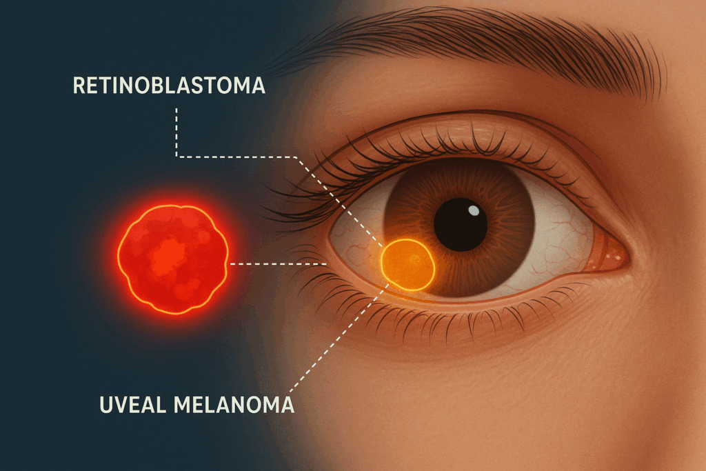
Early detection of eye tumors such as retinoblastoma, uveal melanoma, and other ocular conditions is critical for effective treatment and improved patient outcomes. Achieving this requires precise and meticulously labeled imaging data—something that 𝗠𝗮𝗿𝘁𝗲𝗰𝗸 𝗦𝗼𝗹𝘂𝘁𝗶𝗼𝗻𝘀 specializes in.
By partnering with 𝗠𝗮𝗿𝘁𝗲𝗸 𝗦𝗼𝗹𝘂𝘁𝗶𝗼𝗻𝘀, healthcare and research teams can enhance their AI-driven diagnostic capabilities with high-quality annotation and labeling services tailored specifically for ocular imaging. Here’s how we make a difference:
✅ Pixel-Level Precision: Every B-scan, OCT, and fundus image is meticulously annotated, ensuring that even the smallest features of tumors are accurately captured.
✅ DICOM-Compliant Labeling: Our annotations are fully compatible with standard medical imaging formats, making integration into clinical workflows seamless.
✅ Specialized Feature Marking: We provide detailed labeling for critical indicators such as choroidal thickness and calcification, which are essential for early tumor identification.
✅ Reliable Even with Limited Samples: Our expert team can deliver precise labeling even when sample sizes are small, supporting AI models in challenging scenarios.
✅ Augmented Quality Assurance: Every annotation goes through rigorous quality checks to guarantee accuracy and consistency, giving oncology teams complete confidence in the data.
With 𝗠𝗮𝗿𝘁𝗲𝗸 𝗦𝗼𝗹𝘂𝘁𝗶𝗼𝗻𝘀, AI models for ocular tumor detection become smarter, faster, and safer—helping clinicians detect conditions earlier and make informed treatment decisions.