Ophthalmology is the study and treatment of eye-related conditions and diseases. In the field of ophthalmology, various imaging techniques are used to capture eye images which then aid in the diagnosis, treatment planning, and eye health monitoring. These images can include:
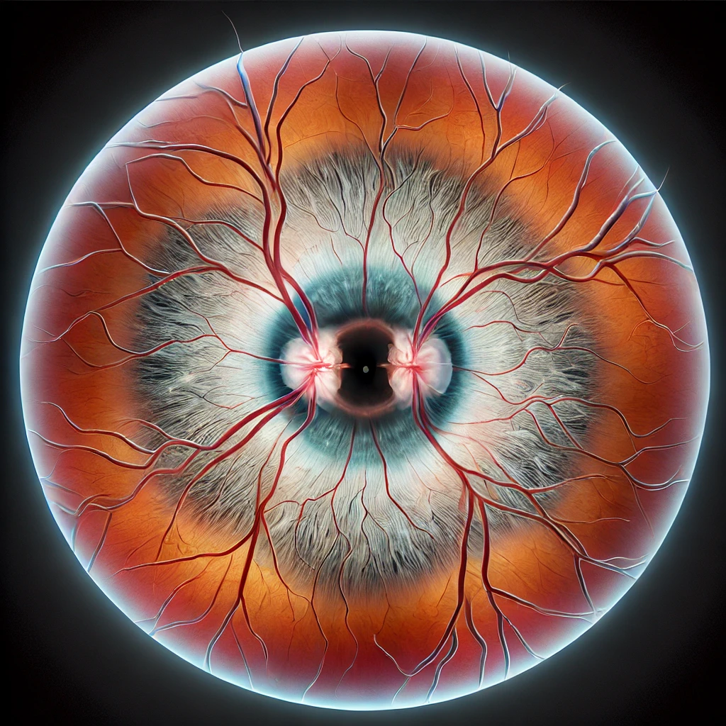
- Fundus Photography
It captures detailed images of the back of the eye, including areas like the retina, optic nerve, and blood vessels. Annotations in these images may involve marking specific features or abnormalities for diagnostic purposes.
- Optical Coherence Tomography (OCT)
OCT provides cross-sectional images of the eye’s layers, which help in the diagnosis of conditions like macular degeneration and glaucoma. Annotations may focus on identifying specific layers or abnormalities.
- Fluorescein Angiography
This imaging Involves injecting a dye into the bloodstream which then highlights the blood vessels in the eye. Annotations are applied to trace the movement of the dye and identify abnormalities for better diagnosis.
- Corneal Topography
This is used to map the surface of the cornea for conditions like astigmatism and keratoconus. Annotations mark irregularities or changes in corneal shape which is crucial.