๐—ง๐˜‚๐—บ๐—ผ๐—ฟ ๐—ฆ๐˜๐—ฎ๐—ด๐—ถ๐—ป๐—ด ๐—ง๐—ต๐—ฎ๐˜ ๐—ฆ๐˜‚๐—ฝ๐—ฝ๐—ผ๐—ฟ๐˜๐˜€ ๐—ฆ๐—บ๐—ฎ๐—ฟ๐˜๐—ฒ๐—ฟ ๐—–๐—ฎ๐—ป๐—ฐ๐—ฒ๐—ฟ ๐—”๐—œ

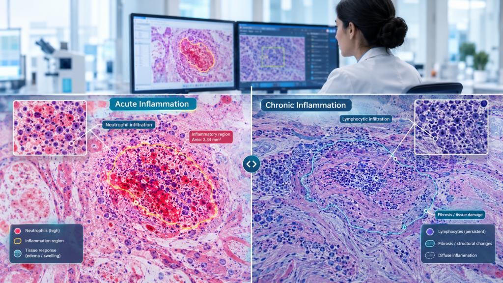
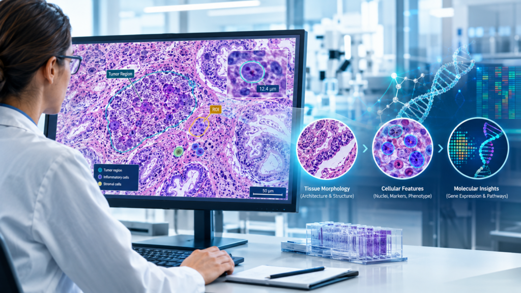
Accurate tumor staging depends on precise identification of the features that influence diagnosis, prognosis, and treatment planning. ๐—ž๐—ฒ๐˜† ๐—ณ๐—ฒ๐—ฎ๐˜๐˜‚๐—ฟ๐—ฒ๐˜€ ๐˜๐—ต๐—ฎ๐˜ ๐—บ๐—ฎ๐˜๐˜๐—ฒ๐—ฟ:โœ… Invasion depth โ€“ Identify tumor penetration across tissue layersโœ… Tumor margins โ€“ Distinguish clear and involved marginsโœ… Structural boundaries โ€“ Precisely delineate tumor extent and spreadโœ… Staging features โ€“ Support consistent clinical classification ๐—›๐—ผ๐˜„ …

๐—ง๐˜‚๐—บ๐—ผ๐—ฟ ๐—ฆ๐˜๐—ฎ๐—ด๐—ถ๐—ป๐—ด ๐—ง๐—ต๐—ฎ๐˜ ๐—ฆ๐˜‚๐—ฝ๐—ฝ๐—ผ๐—ฟ๐˜๐˜€ ๐—ฆ๐—บ๐—ฎ๐—ฟ๐˜๐—ฒ๐—ฟ ๐—–๐—ฎ๐—ป๐—ฐ๐—ฒ๐—ฟ ๐—”๐—œ Read More »

๐—ฃ๐—ฟ๐—ฒ๐—ฐ๐—ถ๐˜€๐—ถ๐—ผ๐—ป ๐—ถ๐—ป ๐—ง๐˜‚๐—บ๐—ผ๐—ฟ ๐—š๐—ฟ๐—ฎ๐—ฑ๐—ถ๐—ป๐—ด: ๐—ฆ๐˜‚๐—ฝ๐—ฝ๐—ผ๐—ฟ๐˜๐—ถ๐—ป๐—ด ๐—•๐—ฒ๐˜๐˜๐—ฒ๐—ฟ ๐—–๐—ฎ๐—ป๐—ฐ๐—ฒ๐—ฟ ๐—–๐—ฎ๐—ฟ๐—ฒ

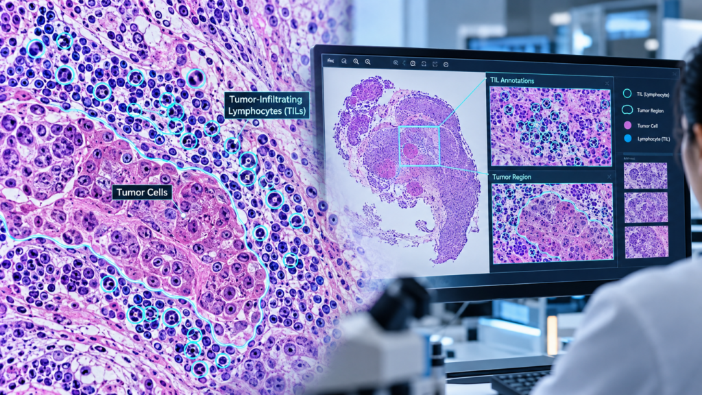
Accurate tumor grading plays an important role in understanding tumor aggressiveness and supporting clinical decision-making. โœ… Low-Grade Tumors โ€“ Typically slower-growing and less aggressive.โœ… High-Grade Tumors โ€“ Generally faster-growing and more aggressive.โœ… Precise Identification โ€“ Consistent annotation helps distinguish important tumor characteristics.โœ… Pathology Annotation โ€“ Supports accurate marking and classification of relevant regions in pathology …

๐—ฃ๐—ฟ๐—ฒ๐—ฐ๐—ถ๐˜€๐—ถ๐—ผ๐—ป ๐—ถ๐—ป ๐—ง๐˜‚๐—บ๐—ผ๐—ฟ ๐—š๐—ฟ๐—ฎ๐—ฑ๐—ถ๐—ป๐—ด: ๐—ฆ๐˜‚๐—ฝ๐—ฝ๐—ผ๐—ฟ๐˜๐—ถ๐—ป๐—ด ๐—•๐—ฒ๐˜๐˜๐—ฒ๐—ฟ ๐—–๐—ฎ๐—ป๐—ฐ๐—ฒ๐—ฟ ๐—–๐—ฎ๐—ฟ๐—ฒ Read More »

๐—–๐—ฎ๐—ป๐—ฐ๐—ฒ๐—ฟ ๐—ฆ๐˜‚๐—ฏ๐˜๐˜†๐—ฝ๐—ถ๐—ป๐—ด ๐—ฆ๐˜๐—ฎ๐—ฟ๐˜๐˜€ ๐˜„๐—ถ๐˜๐—ต ๐—ฃ๐—ฟ๐—ฒ๐—ฐ๐—ถ๐˜€๐—ฒ ๐— ๐—ฒ๐—ฑ๐—ถ๐—ฐ๐—ฎ๐—น ๐—”๐—ป๐—ป๐—ผ๐˜๐—ฎ๐˜๐—ถ๐—ผ๐—ป

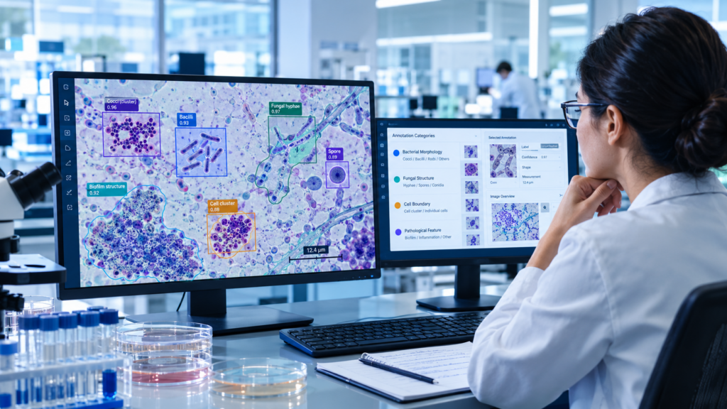
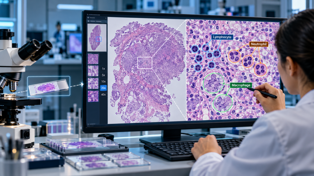
Accurate cancer subtypingโ€”such as distinguishing adenocarcinoma from squamous cell carcinomaโ€”requires more than image classification. Subtle cellular and tissue-level patterns demand specialized pathology expertise and consistent annotation. ๐—ช๐—ต๐˜† ๐—˜๐˜…๐—ฝ๐—ฒ๐—ฟ๐˜ ๐—”๐—ป๐—ป๐—ผ๐˜๐—ฎ๐˜๐—ถ๐—ผ๐—ป ๐— ๐—ฎ๐˜๐˜๐—ฒ๐—ฟ๐˜€:โ€ข Identifies clinically relevant tumor regions and morphological patternsโ€ข Supports consistent cancer subtype classificationโ€ข Helps reduce annotation variability and labeling errorsโ€ข Enables AI models to learn …

๐—–๐—ฎ๐—ป๐—ฐ๐—ฒ๐—ฟ ๐—ฆ๐˜‚๐—ฏ๐˜๐˜†๐—ฝ๐—ถ๐—ป๐—ด ๐—ฆ๐˜๐—ฎ๐—ฟ๐˜๐˜€ ๐˜„๐—ถ๐˜๐—ต ๐—ฃ๐—ฟ๐—ฒ๐—ฐ๐—ถ๐˜€๐—ฒ ๐— ๐—ฒ๐—ฑ๐—ถ๐—ฐ๐—ฎ๐—น ๐—”๐—ป๐—ป๐—ผ๐˜๐—ฎ๐˜๐—ถ๐—ผ๐—ป Read More »

๐—•๐—ฒ๐—ป๐—ถ๐—ด๐—ป ๐˜ƒ๐˜€. ๐— ๐—ฎ๐—น๐—ถ๐—ด๐—ป๐—ฎ๐—ป๐˜ ๐—Ÿ๐—ฒ๐˜€๐—ถ๐—ผ๐—ป ๐—”๐—ป๐—ป๐—ผ๐˜๐—ฎ๐˜๐—ถ๐—ผ๐—ป: ๐—ฃ๐—ฟ๐—ฒ๐—ฐ๐—ถ๐˜€๐—ถ๐—ผ๐—ป ๐—ง๐—ต๐—ฎ๐˜ ๐—ฆ๐˜‚๐—ฝ๐—ฝ๐—ผ๐—ฟ๐˜๐˜€ ๐—ฅ๐—ฒ๐—น๐—ถ๐—ฎ๐—ฏ๐—น๐—ฒ ๐—›๐—ฒ๐—ฎ๐—น๐˜๐—ต๐—ฐ๐—ฎ๐—ฟ๐—ฒ ๐—”๐—œ

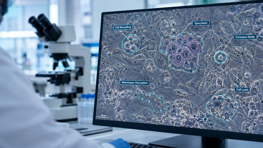
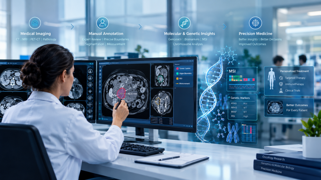
Distinguishing benign from malignant lesions requires more than identifying an abnormalityโ€”it requires clinically accurate and consistent annotation. ๐—ช๐—ต๐˜† ๐—ถ๐˜ ๐—บ๐—ฎ๐˜๐˜๐—ฒ๐—ฟ๐˜€:โœ… Precise lesion boundaries and morphological featuresโœ… Reliable benign/malignant classificationโœ… Better consistency across complex medical imagesโœ… Reduced ambiguity during AI model developmentโœ… Stronger support for AI-assisted clinical decision-making ๐—›๐—ผ๐˜„ ๐— ๐—ฎ๐—ฟ๐—ง๐—ฒ๐—ฐ๐—ธ ๐—ฆ๐—ผ๐—น๐˜‚๐˜๐—ถ๐—ผ๐—ป๐˜€ ๐—ฐ๐—ฎ๐—ป ๐—ต๐—ฒ๐—น๐—ฝ: โœ… Doctor-led medical …

๐—•๐—ฒ๐—ป๐—ถ๐—ด๐—ป ๐˜ƒ๐˜€. ๐— ๐—ฎ๐—น๐—ถ๐—ด๐—ป๐—ฎ๐—ป๐˜ ๐—Ÿ๐—ฒ๐˜€๐—ถ๐—ผ๐—ป ๐—”๐—ป๐—ป๐—ผ๐˜๐—ฎ๐˜๐—ถ๐—ผ๐—ป: ๐—ฃ๐—ฟ๐—ฒ๐—ฐ๐—ถ๐˜€๐—ถ๐—ผ๐—ป ๐—ง๐—ต๐—ฎ๐˜ ๐—ฆ๐˜‚๐—ฝ๐—ฝ๐—ผ๐—ฟ๐˜๐˜€ ๐—ฅ๐—ฒ๐—น๐—ถ๐—ฎ๐—ฏ๐—น๐—ฒ ๐—›๐—ฒ๐—ฎ๐—น๐˜๐—ต๐—ฐ๐—ฎ๐—ฟ๐—ฒ ๐—”๐—œ Read More »

๐—ง๐˜‚๐—บ๐—ผ๐—ฟ ๐˜ƒ๐˜€. ๐—ก๐—ผ๐—ฟ๐—บ๐—ฎ๐—น ๐—ง๐—ถ๐˜€๐˜€๐˜‚๐—ฒ ๐—œ๐—ฑ๐—ฒ๐—ป๐˜๐—ถ๐—ณ๐—ถ๐—ฐ๐—ฎ๐˜๐—ถ๐—ผ๐—ป: ๐—ช๐—ต๐—ฒ๐—ฟ๐—ฒ ๐— ๐—ฒ๐—ฑ๐—ถ๐—ฐ๐—ฎ๐—น ๐—”๐—œ ๐— ๐—ฒ๐—ฒ๐˜๐˜€ ๐—–๐—น๐—ถ๐—ป๐—ถ๐—ฐ๐—ฎ๐—น ๐—ฃ๐—ฟ๐—ฒ๐—ฐ๐—ถ๐˜€๐—ถ๐—ผ๐—ป

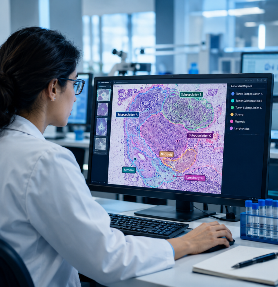
In pathology AI, distinguishing tumor tissue from normal tissue is critical for developing reliable and clinically meaningful solutions. Even small cellular differences can influence diagnosis, treatment planning, and research outcomes. ๐—ช๐—ต๐˜† ๐—ฎ๐—ฐ๐—ฐ๐˜‚๐—ฟ๐—ฎ๐˜๐—ฒ ๐—ฎ๐—ป๐—ป๐—ผ๐˜๐—ฎ๐˜๐—ถ๐—ผ๐—ป ๐—บ๐—ฎ๐˜๐˜๐—ฒ๐—ฟ๐˜€:โ€ข Clear identification of tumor regions and tissue boundariesโ€ข Consistent classification across complex pathology imagesโ€ข Better support for cancer detection and researchโ€ข …

๐—ง๐˜‚๐—บ๐—ผ๐—ฟ ๐˜ƒ๐˜€. ๐—ก๐—ผ๐—ฟ๐—บ๐—ฎ๐—น ๐—ง๐—ถ๐˜€๐˜€๐˜‚๐—ฒ ๐—œ๐—ฑ๐—ฒ๐—ป๐˜๐—ถ๐—ณ๐—ถ๐—ฐ๐—ฎ๐˜๐—ถ๐—ผ๐—ป: ๐—ช๐—ต๐—ฒ๐—ฟ๐—ฒ ๐— ๐—ฒ๐—ฑ๐—ถ๐—ฐ๐—ฎ๐—น ๐—”๐—œ ๐— ๐—ฒ๐—ฒ๐˜๐˜€ ๐—–๐—น๐—ถ๐—ป๐—ถ๐—ฐ๐—ฎ๐—น ๐—ฃ๐—ฟ๐—ฒ๐—ฐ๐—ถ๐˜€๐—ถ๐—ผ๐—ป Read More »

๐—•๐—ฟ๐—ถ๐—ฑ๐—ด๐—ถ๐—ป๐—ด ๐—”๐—œ ๐—ฎ๐—ป๐—ฑ ๐—–๐—น๐—ถ๐—ป๐—ถ๐—ฐ๐—ฎ๐—น ๐—˜๐˜…๐—ฝ๐—ฒ๐—ฟ๐˜๐—ถ๐˜€๐—ฒ ๐—ถ๐—ป ๐—–๐—ฎ๐—ฟ๐—ฑ๐—ถ๐—ผ๐—น๐—ผ๐—ด๐˜†

Cardiology AI needs more than advanced algorithmsโ€”it needs clinically accurate, expert-validated annotations to recognize what truly matters in medical images. At MarTeck Solutions, our medical annotation and labeling services combine clinical expertise with structured, quality-focused workflows to support the development and validation of cardiology AI. 1. Expert-Led Annotation: Cardiologists and trained medical annotators accurately label …

๐—•๐—ฟ๐—ถ๐—ฑ๐—ด๐—ถ๐—ป๐—ด ๐—”๐—œ ๐—ฎ๐—ป๐—ฑ ๐—–๐—น๐—ถ๐—ป๐—ถ๐—ฐ๐—ฎ๐—น ๐—˜๐˜…๐—ฝ๐—ฒ๐—ฟ๐˜๐—ถ๐˜€๐—ฒ ๐—ถ๐—ป ๐—–๐—ฎ๐—ฟ๐—ฑ๐—ถ๐—ผ๐—น๐—ผ๐—ด๐˜† Read More »

๐—ฃ๐—ฟ๐—ฒ๐—ฐ๐—ถ๐˜€๐—ถ๐—ผ๐—ป ๐—ง๐—ต๐—ฎ๐˜ ๐—ฃ๐—ผ๐˜„๐—ฒ๐—ฟ๐˜€ ๐—ฆ๐—บ๐—ฎ๐—ฟ๐˜๐—ฒ๐—ฟ ๐—–๐—ฎ๐—ฟ๐—ฑ๐—ถ๐—ฎ๐—ฐ ๐—–๐—ฎ๐—ฟ๐—ฒ

Accurate medical annotation plays a critical role in advancing AI for cardiovascular care. In 2D Echocardiography, precise identification of cardiac structures and abnormalities can help create more reliable AI-driven insights. How MarTeck Solutions Helps: 1. Expert-Led 2D Echo Annotation โ€“ Precise labeling of cardiac structures, chambers, valves, and regions of interest.2. Pathology-Focused Markings โ€“ Support …

๐—ฃ๐—ฟ๐—ฒ๐—ฐ๐—ถ๐˜€๐—ถ๐—ผ๐—ป ๐—ง๐—ต๐—ฎ๐˜ ๐—ฃ๐—ผ๐˜„๐—ฒ๐—ฟ๐˜€ ๐—ฆ๐—บ๐—ฎ๐—ฟ๐˜๐—ฒ๐—ฟ ๐—–๐—ฎ๐—ฟ๐—ฑ๐—ถ๐—ฎ๐—ฐ ๐—–๐—ฎ๐—ฟ๐—ฒ Read More »

Accurate Cardiac Chamber Detection Starts with Precision Annotation

Reliable cardiac AI begins with one essential foundation: accurate medical annotation and labeling. In echocardiography, cardiac chambers such as the Left Atrium (LA), Right Atrium (RA), Left Ventricle (LV), and Right Ventricle (RV) can vary significantly across views, frames, and patient anatomies. Precise annotation helps AI systems learn these structures with greater consistency. Why precision …

Accurate Cardiac Chamber Detection Starts with Precision Annotation Read More »

Multi-Ethnic Retinal Annotation: Building Fairer AI in Ophthalmology

AI in eye care should perform reliably across diverse populations. That starts with representative, clinically accurate retinal image annotation. Why diversity matters:1. Retinal appearance can vary across populations due to pigmentation and anatomical differences.2. Limited population representation can contribute to inconsistent AI performance.3. Inclusive annotation supports more robust and equitable ophthalmic AI. How MarTeck Solutions …

Multi-Ethnic Retinal Annotation: Building Fairer AI in Ophthalmology Read More »

AI-Powered Uveitis Detection Starts with Precise Medical Annotation

Uveitis can present with subtle inflammatory changes, making accurate identification and monitoring challenging. Multimodal ocular imagingโ€”including fundus photography, fluorescein angiography, and OCTโ€”provides complementary information for evaluating inflammation and retinal changes. Key annotation areas include: 1. Inflammation & Lesion Annotation โ€“ Precisely identify inflammatory regions, lesions, fluid accumulation, and other relevant ocular abnormalities.2. Multimodal Image Labeling …

AI-Powered Uveitis Detection Starts with Precise Medical Annotation Read More »